

Bloodless incision
Radiowave surgery is a great adjunct for any cosmetic practitioner, or for those who perform cutaneous surgery. The basis of radiosurgery is that standard household current is converted to radiowaves at the effective (and patented) level of 4.0 MHz. This is entirely different from standard electrosurgery or Bovie machines, which function in the same manner as a soldering iron. The electrode tip provides the resistance and is heated. Anyone with significant surgical experience can attest to the tungsten electrodes becoming red hot and/or melting with use. This may be sufficient for some modalities, but for cosmetic skin incision it provides increased lateral thermal damage and char, which lead to a compromised scar. Radiowave surgery, on the other hand, is different. Rather than the electrode tip providing the resistance, the tissue provides the resistance and the tip does not get hot. The radiowaves cause intracellular water to boil in a process known as intracellular volatilisation, and the tissues separate with very little blood loss. Simultaneous incision and cauterisation is a tremendous surgical advantage.
Bloodless incision can also be performed with the CO2 laser, which produces light with a wavelength of 10.6 nm in the infrared (invisible) range of the electromagnetic spectrum. The radiant energy produced by the CO2 laser is strongly absorbed by water and by all biologic tissues with a high water content, providing an excellent target for vaporisation. When a small spot size of 0.2 mm is used, the CO2 laser is an excellent incision tool. When larger spot sizes are used, such as the 3 mm handpiece, excellent reduction through ablation and vaporisation is produced.
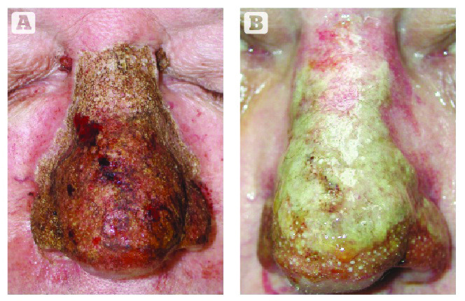
Figure 5 Healing is similar to other skin resurfacing procedures and open wound treatment with petrolatum. (A) Patient immediately after excision and (B) 72 hours post-treatment
Although the CO2 laser with a large spot size is great for reducing tissue, it becomes difficult to debulk thickened tissue as desiccated char forms and reduces the ability of the laser to vaporise. This would be like attempting to reduce a large piece of wood with very fine sandpaper; although it is abrasive, it becomes clogged and inefficient. For gross debulking, the Ellman 4.0 MHz radiowave device is invaluable. Specialised rhinophyma electrodes (Figure 2) are used as a ‘cheese wire’ modality and glide through the phymatous tissue as deeply as the operator desires, with bloodless precision. The bleeding that does occur at the base of the lesion can be treated with the CO2 laser in the defocused mode (holding the handpiece further away from the tissue to enlarge the spot size), or by using the ball electrode with the radiowave system. The ball is used on a pure coagulation setting and provides rapid and effective cauterisation. The author has used both modalities singularly with great success, but prefers to use them in combination for a more effective treatment.
Although rhinophyma sculpting can be performed under local anaesthesia, the author prefers IV sedation as the surgeon is producing a lot of heat and smoke directly into the patient’s face.
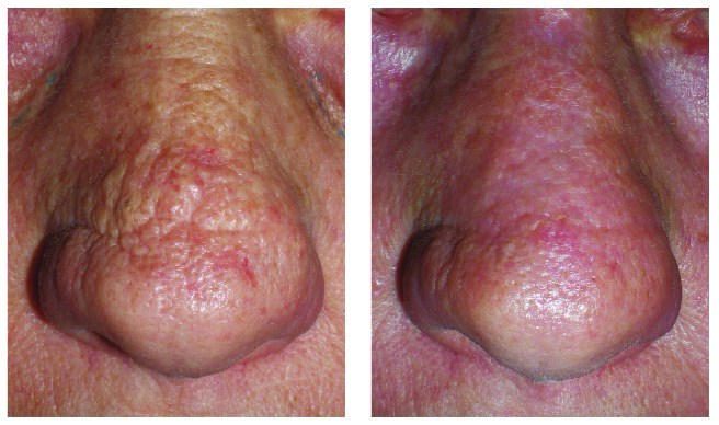
Figure 6 This patient was treated with laser and radiosurgery for minor rhinophyma changes on the nasal bridge
Procedure end-points
The biggest challenge for the novice surgeon is how much tissue can safely be ablated. For those surgeons experienced in operating on the nose, the end-point is usually apparent and the indicators would be a change in the quality of abnormal phymatous tissue to more normal dermis. Other end-point indicators are the residual thickness and dimension of the treated area; that is, the surgeon would stop ablating when the nasal anatomy begins to approximate normal dimensions. It is paramount to remember that when using ablative modalities such as electrosurgery, radiosurgery, laser or ice, there will always be a component of lateral tissue damage that makes the treatment depth deeper than one can see with the naked eye. The surgeon may be removing 100 μm with the laser, but the wound may extend to 125 μm. This is obviously less of a problem with scalpel debulking and similar treatments. The advantages of bloodless incision and debulking without bleeding are huge; less blood means more accurate surgery, less bruising, faster healing, and less postoperative pain.
Mild-to-moderate rhinophyma

Figure 7 This patient was treated with laser and radiosurgery for minor rhinophyma changes on the nasal tip
Mild-to-moderate surgical treatment is essentially simple skin resurfacing. Ablative lasers (CO2, Er:YAG) can be used to ‘burn and wipe’ layers of affected skin much in the same way we treat other areas of the face for wrinkles and actinic damage. In such cases, using the Lumenis Encore (Lumenis Ltd., Yokneam, Israel) laser at a setting of 100 mJ and a density or 6 (30% overlap) performs well at reducing the hypertrophic layers. This is done with a burn/wipe repeated sequence. It is important to bear in mind that with this, and any type of resurfacing treatment: ‘be conservative, you can always take more’. This should be the mantra of any doctor who treats cosmetic patients. It is important to explain to patients that this treatment is a sculpting procedure and although it is easy to remove tissue, it is impossible to ‘put it back’.
Debulking tissue
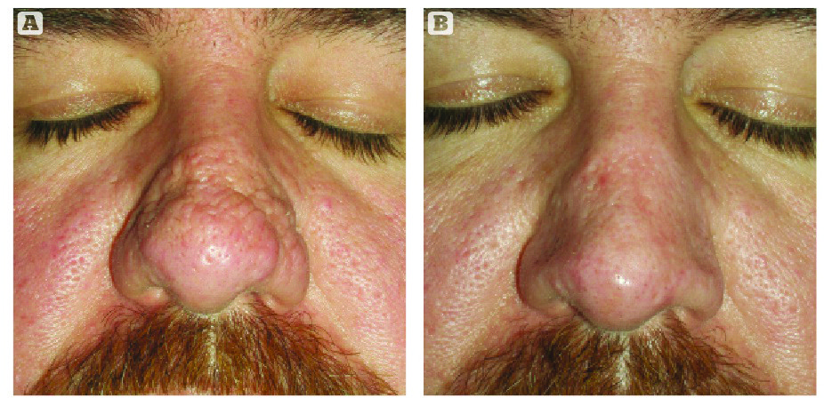
Figure 8 (Above) This patient was treated with laser and radiosurgery for minor rhinophyma changes on the central nose
Larger cases of rhinophyma with bulbous growths require a bigger treatment with a more experienced surgeon. Although there are many ways of debulking this tissue, the author prefers to use the Ellman rhinophyma triangular wire electrode to begin with, and use this in the same manner that a sculptor might remove clay with a curette when working on a bust. These electrodes come at set depths that enable a confluent and homogenous removal of strips of tissue in the same manner as a cheese wire. The author treats one side at a time so that normal tissue dimensions and contours can be approached and then repeated on the other side. This allows the surgeon to use the first side as a standardisation model for the contralateral side (assuming bilaterality).
The setting for the radiowave generator is 10–20 W on ‘cut/coag’, or 30–40 W on the ‘pure coag’ setting. Although this is a coagulative modality, the tissue bed will still bleed and the CO2 laser or Ellman Ball Electrode is used to cauterise the base for haemostasis and fine contouring. When using the Lumenis Encore 3 mm ablative handpiece, a setting of 3–5 W is used. The process is to excise a strip of tissue, then cauterise the base. This is repeated to the desired contour. At this point, ‘fine tuning’ of the tissue contour is completed with the CO2 laser or radiowave ball electrode. Either modality will sculpt and shrink the excess tissue.
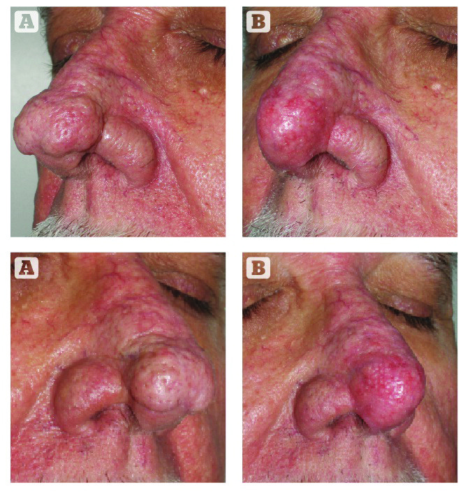
Figure 9 (Below) This patient was treated for advanced rhinophyma with laser and radiosurgery and is shown (A) before and (B) 8 weeks after treatment
Postoperative treatment

Figure 10 This patient was treated for advanced rhinophyma and is shown (A) before and (B) 90 days after treatment with CO2 laser and radiosurgery
Postoperative treatment is very similar to other skin resurfacing procedures, and the author prefers open wound treatment with simple 24-hour coverage with petrolatum or Aquaphor (Beiersdorf AG , Hamburg, Germany) (Figure 5). The tissue will initially appear very dry, brown, and leathery from the desiccation and heat of the procedure. The wound bed will mature to a more pink and granulating state over a number of days, and progress to consistent pink tissue. The erythema can persist for weeks or months, but generally fades faster than similar resurfacing on the cheeks and lower eyelids. Full re‑epithelialisation will occur over 1–14 days. Care after this point is the same as for normal skin.
Selected before and after pictures show minor to moderate rhinophyma treatments (Figures 6–8). Figures 9 and 10 show before and after images of larger rhinophyma cases.
Complications
Complications with rhinophyma include under‑correction with regrowth, over-excision with defects or perforation, burns, scarring, and asymmetry. The author’s experience with rhinophyma treatment has been very positive owing to a conservative approach. This is a condition in which patient happiness is high because the average patient is thrilled to be improved. These have not been ‘picky’ patients; they love what was achieved on a physical, functional, emotional, and physiologic level.
Conclusions
Rhinophyma is an uncommonly seen condition in the average cosmetic surgery practice, but more commonly seen in dermatology practices. It represents the terminal stages of rosacea and has mistakenly been associated with excessive alcohol consumption. Early and minor stages of rhinophyma can be controlled by rosacea medication and trigger avoidance. Moderate and advanced stages require surgical treatment. Haemostatic ablative technologies are the author’s preference for controlled surgical precision and treatment.
The techniques described have proven safe and effective for the functional and aesthetic treatment of rhinophyma.

