

It is important to evaluate each patient individually to assess where the greatest enhancement is needed. Some patients require more volume over the medial cheek, and others over the lateral cheek area. This evaluation for treatment must be performed by assessing the patient from anterior, lateral, and three quarter views. The anterior view is ideal to evaluate where light and shadows fall on the face, whereas the lateral and three quarter aspects are ideal to evaluate malar projection and the ogee curve. Only then, with all aspects in mind as discussed, can one formulate a treatment plan and areas for injection.
Malar enhancement lifts the tear trough in the majority of cases, and further improves overall facial proportions, including infraorbital shadows.
Treatment of the mid-face should not be seen as only improving facial profile and enhancing cheek projection, but should be strongly considered for enhancing the infraorbital region. In a patient with loss of fat in the mid-face, the shadows of the tear trough and nasojugal fold are visible, as well as a lack of the ogee curve on a three quarter view of the patient. This indication can ideally be treated with a high viscosity volumising dermal filler. The needle technique for this specific indication can be used in a three-point technique along the cheekbone, placing three boluses according to the amount of volume needed in the area to be treated. The line of placement of the boluses is 1 cm below the inferior orbital rim and according to patient-specific evaluation. An amount of 0.5–2 ml per side may be needed for enhancement. The fanning technique may also be used to enhance the mid-face, but may result in an increased risk for bruising owing to the multiple movements made with the sharp needle in a fanning technique.
The fanning technique is more ideally suited to the use of a cannula for mid-facial enhancement. The insertion point for the cannula may be at a medial or lateral aspect depending on the injector’s choice and the area requiring the most enhancement. The medial insertion point is 1.5 cm lateral to the nasal alae, and the lateral insertion point is 2 cm lateral and 2 cm inferior to the lateral canthus. Cannula size is selected according to the dermal filler used, and may vary from 22 G to 27 G for this indication. The technique used with the cannula is fanning into the desired region of enhancement.
Both needle and cannula techniques are popular for mid-facial enhancement among practitioners.
Possible complications
Treatment of the periorbital area is very rewarding in most cases, but practitioners should be aware of the possible complications. The majority of complications are seen with filling of the tear trough, and include:
-
Incorrect treatment plan in which malar enhancement was not considered. Opting for malar enhancement can often prevent the swelling and periorbital oedema associated with tear trough filling
-
Too much product can cause a blue undertone and worm-like appearance under the eye. This should be treated with hyaluronidase (if available in country of practice)
-
Incorrect placement of the filler owing to incorrect evaluation and inspection during treatment
-
Bruising and haematoma can be reduced by using cannulae
-
The risk of embolisation appears to be significantly less with fillers compared with autologous fat.
The above tear trough complications can be avoided by clearly distinguishing between tear trough filling exclusively and cheek lifting to avoid incorrect placement of product. The use of a low or medium viscosity hyaluronic acid filler is best as high viscosity products are more likely to cause oedema. Ensure that the filler is not placed too superficially to avoid a blue discolouration.
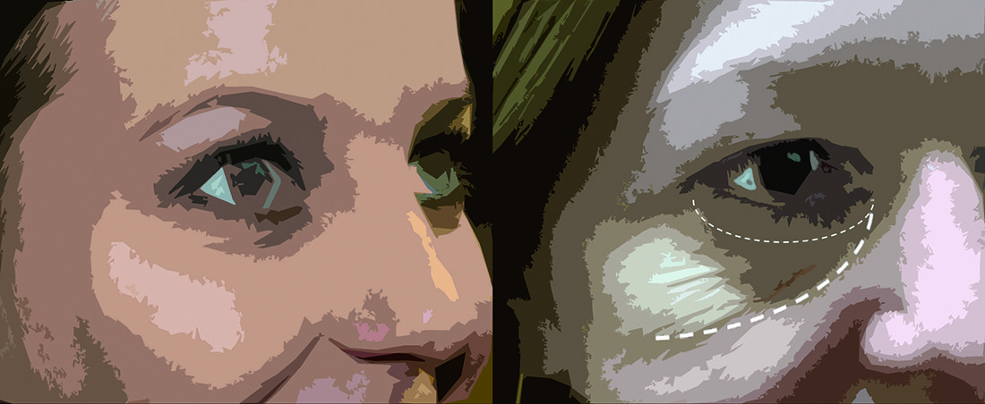
Figure 5 A typical patient who only needs tear trough enhancement compared with a more mature patient who will benefit from malar enhancement
Bruising and haematoma can be reduced by carefully evaluating the location of vessels. Should this occur, immediate direct pressure for 2 minutes will minimise the unsightly appearance. Cooling devices used on the skin before injection are helpful in areas prone to bruising (as is the periorbital area) by enhancing the vasoconstrictive effect.
The feared risk of intravenous placement of fillers should be reduced with a good anatomical knowledge and a careful evaluation of vessels under optimum lighting conditions. It is also essential to monitor any reaction of the skin during injection. A blanching (whitening) of the skin indicates intravascular placement and this is often associated with a sudden intense pain felt by the patient. Injection should be stopped immediately and the appropriate protocol for this complication followed. Treatment with Hyalase (if available) and local massaging should be initiated. The patient will also need daily follow-up for 1 week to monitor the skin and treat accordingly.
Conclusions
Periorbital rejuvenation will always remain a significant demand from patients, as this is the core of their face and expression. As aesthetic doctors, it is paramount to keep updated with high quality scientific teachings, articles and workshops
With regard to periorbital rejuvenation, it is even more important to have an in-depth knowledge of anatomy owing to the vital structures and multiple vascular structures in this area. Knowledge of the pathological changes of ageing in the periorbital and mid-face regions helps one to understand and formulate a better treatment plan. All technical knowledge should be complemented with an holistic view of the patient and his/her individual needs. This will ensure that we complement the patient’s unique lifestyle and appearance.

