

In the world of aesthetic medicine, there has been a recent increase in demand for breast augmentation and soft tissue shape correction procedures to the breast. The number of patients who wish to enlarge the breast soft tissues or improve the shape of their breast is the reason why surgeons continually search new minimally-invasive methods.
Currently, growing demand for Aquafilling Breast Augmentation® (ABA) is being observed in the aesthetic market. It is a minimally-invasive procedure performed under local anaesthesia, with a short and favourable postoperative period, through the application of the hydrophilic gel Aquafilling®. It is a volumising filler comprising 97–98% physiological solution of sodium chloride and 2–3% linear polyamide.
The advantages of Aquafilling® are its high biological adaptability, caused by the content of physiological solution in the filler, and its expressed plasticity as a result of the spatial structure of the gel. The gel is injected in the retromammary region using a 16–18 G implantation needle or cannula (sub-mammary or upper lateral access). After application of the gel according to the methodology of ABA, the patient can return to her social life just 1 day after treatment.
Study objective
In the present study, the authors aimed to evaluate the possibilities of ultrasonographic monitoring of mammary glands of patients who underwent ABA.
The study tasks concerned the following:
-
To examine the sonographic structure of mammary glands after ABA
-
To evaluate the dynamics of sonographic pictures of mammary glands with different terms after implantation
-
To examine the characteristics of blood flow in the mammary gland tissues after ABA
-
To evaluate identification possibilities of the development of any abnormal processes in the mammary gland tissues in patients who underwent ABA.
The indications for ABA were correction of hypomastia, ptosis and asymmetry of breast soft tissues.
Materials and methods
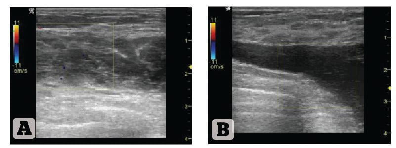
Figure 1 Ultrasound structure of the right mammary gland. (A) Patient 1, 45 years old, 9 years after ABA; (B) Patient 2, 26 years old, 2 years after ABA
Seventeen patients aged 26–45 years were enrolled into the study. The subjects underwent breast augmentation with application of hydrophilic gel Aquafilling®. The volume of the injected gel amounted to 150–250 ml in each breast’s soft tissue. The volume of 250 ml was injected in two stages, as the recommended volume of gel for each procedure is 150 ml.
The follow-up period ranged from 2 to 9 years. The investigation was conducted using ultrasound device LOGIQ-P5 at the medical centre ‘La Vita Sana’, Kharkiv, Ukraine, from 24 April 2013 till 20 May 2013.
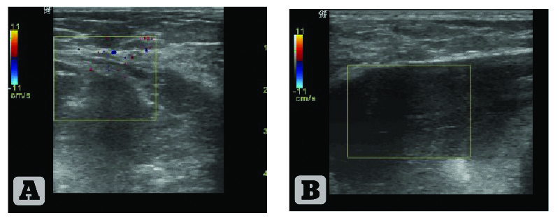
Figure 2 Ultrasound structure of the left mammary gland. (A) Patient 1, 45 years old, 9 years after ABA; (B) Patient 2, 26 years old, 2 years after ABA
During the long-term postoperative period the following indices were examined in all patients:
-
Parenchyma of mammary glands
-
Ligaments
-
Blood flow in the tissues, adhered to the implant
-
Axillary lymph nodes.
A particular focus was given to investigation of the implant structure, its homogeneity, and character of perifocal manifestations in the surrounding tissues.
Discussion
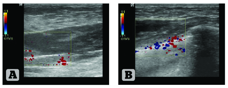
Figure 3 Ultrasound structure and blood flow of the right mammary gland. (A) Patient 1, 45 years old, 9 years after ABA; (B) Patient 2, 26 years old, 2 years after ABA
In all cases, the gel was observed in the form of homogeneous hypoechoic mass placed in the retromammary space. There were no signs of gel migration. A very thin capsule around the implant can be seen, which densely adheres to the walls of the retromammary space. Implant invasion with connective tissue was not recorded, as well as availability of calcificates. Nothing abnormal with regard to blood flow in the tissues of the mammary glands during Doppler sonography was detected. Axillary glands did not increased.
Figures 1–4 show comparative data of ultrasonographic pictures of mammary glands in two patients with implantation terms of 2 and 9 years after ABA. No vital distinctions in the structure of the mammary glands and surrounding tissues were detected in these patients.
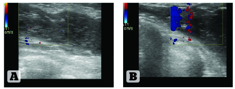
Figure 4 Ultrasound structure and blood flow of the left mammary gland. (A) Patient 1, 45 years old, 9 years after ABA; (B) Patient 2, 26 years old, 2 years after ABA
Conclusions
Ultrasound sonography can be considered a screening method in the monitoring of mammary glands in the patients after Aquafilling® Breast Augmentation (ABA). No changes to the space structure of the implant injected into the retromammary space, or any other degenerate processes in the tissues of the mammary glands, were recorded during the long-term follow-up period. Taking into consideration a distinct differentiation between the tissues of mammary glands and the implant, ultrasound sonography makes it possible to monitor different abnormal processes in the mammary gland tissues at the early stages of development, unobstructed by gel.

