

Melasma
Melasma is a commonly-acquired, localised hyperpigmentation disorder characterised by irregular brown or grey–brown macules and patches with well-defined margins that occur symmetrically on sun-exposed areas of the body, usually the face19–20. This condition typically affects women of child-bearing age, and solar and UV exposure are the best known aetiologic factors. Histological and immunohistochemical studies have shown that melasma skin presents features of prominent solar damaged skin. UV irradiation is known to increase the synthesis of alpha-melanocyte-stimulating hormone (α-MSH) and adrenocorticotropic hormone (ACTH) derived from proopiomelanocortin (POMC) in keratinocytes. These peptides lead to the proliferation of melanocytes, as well as an increase in melanin synthesis via stimulation of tyrosinase activity and tyrosinase-related protein-1 (TRP-1)21.
One study has shown that melasma is characterised by alterations in the dermal structures in addition to pigmentary changes, suggesting a role of the dermis in melasma development22. The role of fibroblasts in melasma development has also been suggested. In fact, the over-expression of both stem cell factors (SCFs) from fibroblasts and c-kits has been found: the fibroblast-derived cytokines stimulate proliferation and melanogenesis of melanocytes in culture. Therefore, it is possible that the dermal inflammation induced by an accumulation of UV irradiation may be associated with the activation of fibroblasts, which results in the upregulation of SCFs in dermal melasma, leading to increased melanogenesis22. Recent data has also shown that melasma lesions have greater vascularisation than normal perilesional skin. An increased expression of the vascular endothelial growth factor (VEGF) in keratinocytes has been suggested as a significant angiogenic factor for altered vessels in melasma23; therefore, the network of cellular interactions between keratinocytes, fibroblasts and perhaps vasculatures and melanocytes during chronic sun exposure may play an important role in the development of melasma, working in conjunction to stimulate melanocytes, resulting in epidermal hyperpigmentation.
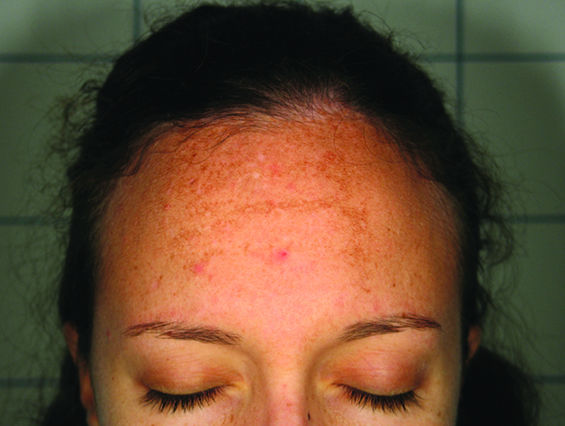
Figure 4 A patient with a facial melasma with the pigment almost along forehead wrinkles
Melasma can be classified depending on the site of the lesions (craniofacial, malar, mandibular), histological depth of the pigmentation (epidermal, dermal, mixed), and appearance under the Wood’s lamp (epidermal, dermal, mixed, indeterminate):
- ■ Epidermal:Light brown with an enhancement of pigmentation under the Wood’s lamp. Histologically characterised by a melanin increase in the basal, suprabasal and stratum corneum layers
- ■ Dermal: Ashen or blue–grey with no enhancement of pigmentation under the Wood’s lamp. Histologically there is a predominance of melanophages in the superficial and deep dermis
- ■ Mixed: Dark brown with enhancement of pigmentation under the Wood’s lamp in some areas only
- ■ Indeterminate: Not detected under the Wood’s lamp.
The best therapeutic results are normally achieved in epidermal melasma24–25. The laser to be used must be selectively chosen and should generally be used in cases in which there is proven resistance to conventional treatments.
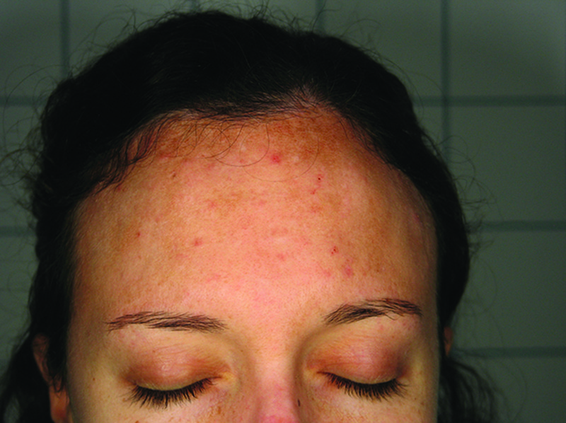
Figure 5 After three intense pulsed light (IPL) (Photosilk Plus, DEKA Laser, Florence, Italy) sessions and one session of the short-pulse alexandrite laser (Synchro Repla:Y, DEKA Laser, Florence, Italy) hyperpigmentation is reduced
Q-switched lasers
In the past, attempts to treat melasma with lasers that targeted melanin, such as the QS ruby laser (694 nm), short-pulsed green dye laser (504–510 nm), QS neodymium laser (1064 nm), and argon laser (514 nm), yielded disappointing results. For example, in a randomised controlled trial conducted by Wattanakrai et al26, 22 patients with dermal or mixed melasma were treated with the same laser at a fluence of 3–3.8 J/cm2 for five sessions at 1-week intervals. The treatment was combined with 2% hydroquinone and compared with hydroquinone alone. There was a 92.5% improvement, which was significant compared with the control group. However, 13.6% of patients developed faint, spotty hypopigmentation that improved during follow-up. Furthermore, 18% of patients developed rebound hyperpigmentation, and all patients had a recurrence of melasma.
Jeong et al27 compared the clinical efficacy and adverse effects of the low fluence QS Nd:YAG (1064 nm) laser when performed before and after treatment with topical triple-combination (TC) creams using a split-face cross-over design in 13 patients with melasma. They used a collimated 5–7 ns pulse width, 7 mm spot size, and a fluence of 1.6–2.0 J/cm2. Weekly sessions were carried out for 8 weeks. The laser was compared with pre- or post-treatment TC cream. The authors found that pre-treatment with TC creams was more effective as this decreases melanin production before laser injury; therefore the risk of PIH is reduced and the melasma improves. If TC cream is used after laser treatment, the melanin is produced at full capacity with a higher risk of PIH and less improvement in the melasma. Consequently, the authors recommend medical treatment for hyperpigmentation for at least 8 weeks before laser treatment in order to achieve optimal results.
Kauvar28 assessed the safety and efficacy of a procedure combining microdermabrasion, a topical regimen, and low fluence QS Nd:YAG laser treatment in 27 female subjects. In particular, low fluence QS Nd:YAG laser treatment of 1.6–2 J/cm2 with 5 mm or 6 mm spot was administered immediately after microdermabrasion. Treatments were repeated at 4-week intervals. Twenty-two subjects (81%) had more than 75% clearance of melasma; 11 subjects (40%) achieved more than 95% clearance. Most subjects showed more than 50% clearance of their melasma 1 month after the first treatment. Side-effects were limited to mild post-treatment erythema, which developed after the microdermabrasion and lasted approximately 30–60 minutes. Remission lasted for at least 6 months.

