

Indications for cervicofacial liposculpture
With an aesthetic view in mind, these indications apply to making the face both more beautiful and youthful. In the young adult, whose skin is firm and elastic, the restoration of a normal cervical chin contour through liposuction must follow the criteria elaborated by Ellenbogen et al3 and Karlin:
- A well delimited mandibular edge
- A visible under-hyoid depression
- A visible thyroid cartilage feature
- A well defined sternocleidomastoid anterior edge
- A cervical chin angle of 105–120°.
In older patients, the rebuilding of a three‑dimensional image conveys the importance and consistency of the existence of fat in any thin face. The sagging of the skin itself is important, and is almost always associated with low cheeks. The quality of the skin will further deteriorate with dehydration.
A normal cervical chin contour ensures not only a perfect facial oval from the front, but also guarantees a better profile. Correction of the under chin and under maxillary ptosis gives better chin definition. Liposuction is also an excellent indication for male patients as they have thicker skin and fewer wrinkles as a result of facial hair.
Preoperative assessment
Undertaking an initial questionnaire is paramount before any procedure to record the patient’s medication history, and also because it is common to find a poly‑medication regimen among older patients, particularly with regard to anti-coagulants or the prescription of small quantities of aspirin with a preventive cardiovascular aim.
The physician should also pay particular attention to some harmful associations
with lidocaine, such as beta blockers, for example. Should anti-coagulants be taken, the patient should stop taking them 48 hours before the intervention and resume taking them the day after, pending the cardiologist’s agreement
The traditional check-up should investigate platelets, activated partial thromboplastin time (APTT), glycaemia, HIV serodiagnosis, and hepatitis.
Patient preparation
Disinfection is paramount, and the patient should shower with an anti-bacterial soap the day before and on the day of treatment.
Preoperative markings are very important as they provide excellent landmarks of the area to be treated once the hydrotomy is complete. Marking with potassium permanganate in a saturated solution presents two advantages. Firstly it avoids hyperpigmentation at the entry point, and secondly, this marking persists throughout the whole intervention, despite Betadine and draining of the solution on the skin. Marking should be carried out while the patient is in a reclining position. It is essential to clearly define the cervical thickening on the double chin and lower cheeks, which play an essential part in the final aesthetic result. Finally, locate the eventual hollows in order to be sure to suction at this level and avoid worsening any pre‑existing conditions.
Anaesthesia technique
The author uses Klein’s tumescent infiltration technique4 with the following formula:
- 1000 cc physiological serum
- 600 mg xylocaine
- 10 cc sodium bicarbonate at 1.4%
- 1 mg adrenaline.
The infiltration materials include an infiltration cannula with a 2 mm diameter (or spinal needles), and a 10 cc luer lock syringe or infiltration pump.
The author makes a number of 1% adrenalined, xylocaine dermic spots. The Klein solution is slowly injected and remains at the subcutaneous plane. As soon as pain is felt, the area is gently massaged to allow for an homogeneous distribution of the solution and diminish any pain linked to tissue stretching. This
slow tissue stretching associated with the vaso‑constrictive adrenaline action creates a tourniquet at the level of the SACS arteriovenous system, and thus reduces the risk of anaesthetic products infiltrating the intravascular bolus. Skin colour becomes white, proving that the adrenaline has worked.
Infiltration is limited at the top by the orbital edge and within by the nasogenian grooves, and outside by the pre-auricular area. It goes beyond the mandible and spreads to the under chin area as far as the sternum.
The Klein solution infiltration is administered at 400 cc. Once the infiltration is over, wait for at least 30 minutes in order to let the solution work at the tissue level. A cold facial mask is applied with the aim of bettering the vasoconstrictive effect through the cold. The cold diminishes the residual pains linked to the infiltration and through this analgesic action reduces the patient’s stress.
A major hydrotomy results in great adipose tissue tumescence. Quantities of 250–400 cc are usually administered. This also results in being able to safely work on deeper planes and avoids damaging noble elements at the levels of the neck and face.
Surgical technique
Two punctiform incisions are made at the jugal level (i.e. zygomatic) with a n°11 surgical scalpel blade in the lower third of the nasogenian groove, and 2 cm ahead of and within the tragus in the pre-auricular area. Three incisions are made under the chin: under the chin tip; under the mandible; and opposite the branch travelling upward from the inferior maxilla.
Depending on the shape of the neck, the author will decide whether to make an incision at the tip of the cervico–chin angle. In some cases of a closed angle, this additional incision is necessary to allow regular work in a cross-section and obtain a satisfactory retractile cicatricial fibrosis. As Fournier taught, ‘it is necessary to get as close as possible to the default to be treated’ 5. Too distant a treatment with a long cannula risks being unlevel to the cutaneous plane and can damage the vessels or subjacent nerves.
However, Fournier’s entry points do not allow the physician to efficiently treat the lower cheeks. The entry point at the lower third of the nasogenian groove allows for an efficient treatment of the lower cheeks through the tunnelisation crossing at that level.
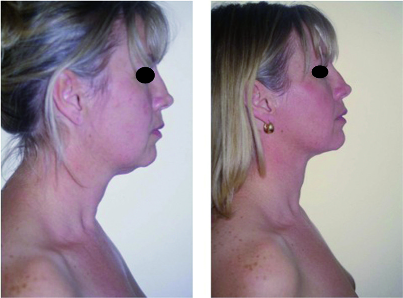
Figure 1. 38-year-old female (left) before and (right) 2 months after treatment
The first step of the operation requires a careful tunnelisation, or cannulation, that is non-aspirated and in cross-sections, with a 2 mm cannula. The free hand is used to stretch the tissues by spreading the thumb and fingers on the skin, limiting a subjacent effraction risk. One hemiface is treated first, making it possible to appreciate the oval of the face that has been redesigned by lipodestruction induced by tunnelisation. The second hemiface is then symmetrically treated.
The second step of the operation comprises a lipoaspiration adapted to the shape of the face. The author will usually use a 1 mm olivary-tipped cannula, which allows a gentle and neat aspiration that is also quite precise — avoiding bleeding — and a 5 or 10 cc luer lock syringe fitted on a blocker. The area under the chin is carefully treated, then a degressive action is applied from the mandible contour to the malar area. Naturally, liposculpture is more important on the mandible contour and insists on thicker areas. Skin sagging traction and pinching make it possible to control the fat resection: elasticity must be retained. The path of the cannula must also be parallel to the cutaneous plane; both entry points made at the jugal level make it possible to cross the tunnelisation planes. The surgeon operates either by ‘touching’ the tissue between thumb and index finger, or by a palpate and roll massage that guides the cannula trajectory and thus allows the surgeon to work on both the surface and at depth at the same time.
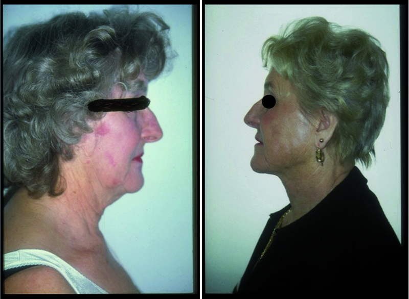
Figure 2. 76-year-old patient (left) before and (right) 1-year post-treatment
The third step of the operation comprises a peripheral lifting with a classic one-hole cannula. This lifting is carried out at each entry hole, allowing treatment to both the neck and the lower cheeks, and guaranteeing a good cutaneous tightening. Incisions are covered with Steri‑Strip.

